The incidence reported in the literature ranges from 0.4-8.4%third molars and in some instances the damage can be permanent. The aim of this case-control study was to investigate the specific risk factors for neurosensory deficits of inferior alveolar nerve (IAN) after third molar extraction.
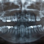
The cases consisted of patients showing neurosensory deficits of the lower lip and mental area after mandibular third molar extraction. The control group consisted of randomly selected patients who had undergone third molar extraction without any subsequent neurosensory symptoms after the procedure. The authors considered a number of predictor variables grouped as demographic, radiographic, and anatomic. The radiographic variables included the impaction depth and angulation of the mandibular third molar. The impaction depth was classified according to the Pell-Gregory classification angulation was classified according to the Winter’s classification. The anatomic variable, were classified according to the Rood and Shehab 7-type classification. The area of paraesthesia was recorded clinically using 2-point discrimination, pin- prick test, and light touch detection, using Semmes- Weinstein monofilaments. Recovery was assessed as decreases in area with dysaesthesia and a complete disappearance and a decrease in discomfort or no additional visits
- 104 cases and 135 controls were included
- Older age and deeper impaction status were significant risk factors (P < .05). Darkening of the roots, deflection of the roots, narrowing of the roots, dark and bifid apexes of the roots, and narrowing of the canal were also significant risk factors.
[table id=33 /]
* Statistically significant
- However, the relatively low positive predictive value renders questionable the predictability of superimposition signs on orthopantomography. In the absence of specific radiographic signs, the risk of neurosensory deficit of the IAN could be negligible.
- The sensory symptoms disappeared after 6 months in 92.3% of the patients and 98.1% showed recovery after 1 year.
The authors concluded
The results of the present study have demonstrated a significant association between several risk factors and neurosensory deficits of the IAN after third molar extraction.
Links
Kim JW, Cha IH, Kim SJ, Kim MR. Which Risk Factors Are Associated With Neurosensory Deficits of Inferior Alveolar Nerve After Mandibular Third Molar Extraction? J Oral Maxillofac Surg. 2012 Aug 15. [Epub ahead of print] PubMed PMID: 22901857

Interesting.
You are really doing a great job with this website. Thanks!
Thank you Allesandro
and thanks also for your comments on some of our previous posts.
The Dental Elf
[…] Dental Elf – 5th Sept 2012- Significant association between several risk factors and Inferior … […]